


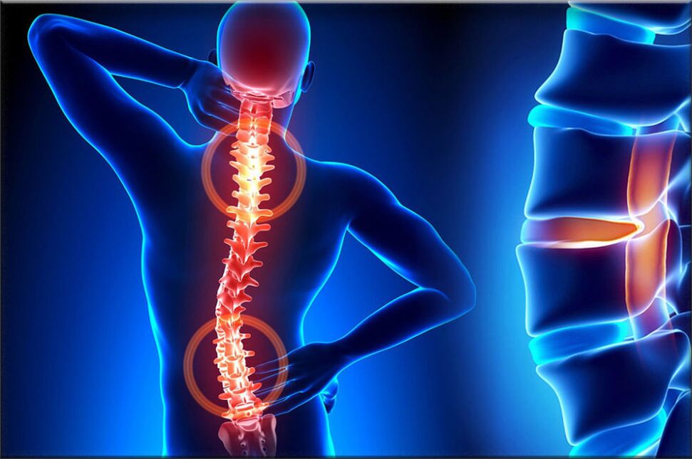
Osteochondrosis is one of the most common diseases of the musculoskeletal system, which manifests itself as a result of a complex of certain dystrophic changes in the cartilage of the vertebra, during this pathological process, the discs of the spine are often affected. The structures, which are the intervertebral cartilage discs, provide flexibility, and also allow the human spine to move, that is, they provide movement.
With osteochondrosis, a number of processes occur that cause degeneration in the vertebral discs, as a result of which they begin to lose elasticity and reduce the degree of flexibility, and at this time the disc itself becomes quite flat. The distance between the two discs decreases, while compressing the nerve endings and blood vessels and causing severe pain. The site of compression of the nerve node begins to swell, which leads to an increase in pain and even greater infringement.
During the development of osteochondrosis, muscle structures and most organs of the body are often involved in this pathological process. This is due to the fact that during the maximum infringement of the neurovascular bundle, blood circulation and motility of muscles and organs are disturbed. For example, the most common osteochondrosis is cervical osteochondrosis, which is accompanied by pain in the back of the head, nausea, dizziness, visual impairment and often tinnitus. This disease has become quite "younger": a century ago, osteochondrosis was a disease of people of gerontological age, and today young people are also susceptible to it.

The most vulnerable category of people is those who have severely impaired metabolism and hormonal levels of the body, as well as people who have disorders of the vascular-venous nature. This is due to the fact that these diseases cause disruption of disk oxygenation. If qualified, timely measures for healing are not taken, then the edges of the affected intervertebral disc, which is compacted, will anatomically protrude beyond the limits of the spinal column, thereby destroying the neurovascular bundles.
Because of this, the patient is at risk of having a herniated disc. The main, significant cause of osteochondrosis is the uneven distribution of the load on the spine, which leads to the fact that the cartilaginous structure changes at points with excessive pressure. The nature of this disease depends on the stage and level of damage to the affected discs. Intervertebral discs change with age, like our hair. Major injuries or fractures of the spine can affect how they function. Casual clothing and certain types of vibration can also accelerate the rate of spinal degeneration. In addition, evidence suggests that smoking increases the rate of spinal degeneration. Scientists have also found a link between family members, highlighting the role of genetics in how quickly change occurs.
The disease can also be triggered by a variety of factors:
- injuries, bruises;
- dystrophy of the spinal muscles;
- stoop and curvature of the spine;
- lifting weights;
- prolonged stay in one position;
- metabolic disease;
- lack of trace elements and vitamins - manganese, magnesium, zinc and vitamins D and F;
- hereditary predisposition;
- physical overload;
- sedentary lifestyle;
- radiation background;
- frostbite;
- congenital dystrophies;
- asymmetrical work of the muscles of the spinal column;
- stress, depression.
These causes of osteochondrosis are just the assumptions of scientists, direct factors that cause the disease, science has not yet found, and we are talking only about risk factors.
First perioddevelopment - characterized by early deployment of the intradiscal nucleus pulposus (nucleus pulposus of the eccentric intervertebral disc, located next to the dorsal part of the vertebra).
Second periodcharacterized by the appearance of instability of the spinal segment. Pathological substrates are represented by the fibrous core of the affected disc with degenerative processes of take-off and fragmentation of the posterior longitudinal ligament, pathological movements between the vertebrae develop.
Third periodthe development of the disease - total damage to the intervertebral disc, with the appearance of a "herniated disc" - dislocation and exit of fragments of the nucleus pulposus outside the intervertebral space.
If the disease has reached the third phase, then here the process of destruction is already irreversible and can lead to profound disability.
Types of osteochondrosis
The evolution of osteochondrosis is slow, with exacerbations caused by spinal injuries, exercise, carrying weights, etc. The clinic depends on the location of the lesion.
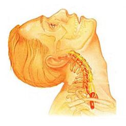
Osteochondrosis of the cervical spinehas local and remote symptoms of advanced forms - with loud root domination, that is, it contributes to the development of severe radicular pain. Symptoms of osteochondrosis in the cervical spine are accompanied by varying degrees of dysfunction, sometimes manifested in a sudden limitation of the mobility of the cervical spine and functional blocks. Headaches can be both pulling and paroxysmal in nature with irradiation to the interscapular region or shoulder region. In the acute period, patients are diagnosed with attacks of pain in the neck, which impede and restrain the movement of the head and neck. In addition to severe discomfort, pain syndrome can be accompanied by dizziness, insomnia, pain, loss of appetite, depression, diseases of the eyes and pharynx.
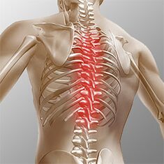
Chest osteochondrosis. . . Clinical manifestations are due to local lesions and processes of destruction of the root structure of the nerve. Thoracic osteochondrosis has a pronounced pain syndrome, which can have a chronic or acute nature of back pain with discomfort in the chest and limited muscle contracture, up to right-verbal muscle atrophy. Chest pain can manifest itself as diffuse, intercostal, and neuralgic. Palpation enhances the axial rotation of the vertebral body. Disorders correspond to the level of root irritation from Thl1 to Thl2, and can manifest themselves as angina pain, reflected in dysfunction of the liver and gastrointestinal tract. Disorders of the genitourinary system and genital area often occur. Patients note sensory disorders such as paresthesias, superficial and deep sensitivity is significantly reduced.
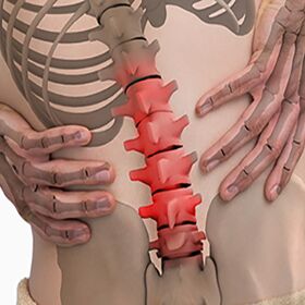
Lumbar osteochondrosis. . . It is characterized by abdominal reflexes and dysfunction of the lower extremities. During the development of neurological disorders, muscle weakness in the legs and pelvic organ dysfunctions can occur. Osteochondrosis is characterized by assessing the damage to the sitting process. The more advanced the stage of development of the lesion of the lumbar vertebrae, the shorter period of time the patient can sit. The lumbar forms are characterized by chronic and acute back pain, spasm of the paravertebral muscles, and myofascial secondary syndrome. The pain radiates to the buttocks and posterior ilium.
Depending on the localization of the pathological process of osteochondrosis, the disease can lead the patient to a violation of superficial sensitivity (tactile, thermal). Also characteristic are changes in reflexes (for example, there is no Achilles reflex), muscle wasting, muscle tone disorders, vegetative manifestations disorders (pallor, redness of the skin, trophic changes in nails, skin hypothermia in the distal extremities), sphincter dysfunctions and sexual dysfunctions.
Clinical picture
Diagnosticsbegins with a complete history and physical examination. The doctor asks questions about the symptoms, how the disease interferes with the patient's daily activities. Also, the specialist is interested in identifying positions and activities that emphasize or reduce the level of pain.
The doctor then examines the patient, checking the position and range of motion in the spine, thereby determining which movements are causing the pain. Skin sensitivity, muscle strength and reflexes are tested equally. Based on the medical history and physical examination, the doctor determines which techniques will help.
Radiography rarely helps with diagnosis, no more than 30% of radiographic images show abnormalities in the early stages of the development of the disease.
However, if the symptoms are severe and the disease is already in its second or third stage, defects in one or more intervertebral discs can be seen in the image. They can be penetrated by osteophytes between the vertebrae and joints.
If additional information is needed, magnetic resonance imaging is prescribed. MRI is used to view the soft tissues of the body. This is useful if the tissue core is absorbing water, or if there are cracks inside the disc. An MRI can show problems in other soft tissues, such as spinal nerves.
Discography can aid in the diagnosis. This examination is performed using a contrast agent, which is appropriately injected into one or more discs. Subsequent viewing on radiography provides useful information about the condition of the discs.
Treatment of osteochondrosis, depending on the varieties
Non-surgical treatment of osteochondrosis
Whenever possible, doctors prefer non-surgical treatment. The most important thing in non-surgical treatment is to relieve pain and other discomfort so that the patient can resume a comfortable standard of living as much as possible.
Doctors rarely prescribe bed rest for patients with osteochondrosis problems. Patients are encouraged to live in natural mobility when pain is not a concern. If symptoms are severe, several days of bed rest may be prescribed.
When the spine is displaced, an elastic belt is sometimes prescribed, which is worn for no more than 2-4 days in order to avoid atrophy of the back muscles.
Osteopathic sessions provide serious relief from osteochondrosis.Osteopathic physiciannot only diagnoses a problem area, but also relieves pain in 1-2 doses, relieves the general condition of the body and "tightens" the visceral organs.
Patients may be prescribed medications to control symptoms and to resume normal activities for a long time. If symptoms continue to restrict the patient's activities, a conventional physician may suggest an epidural steroid injection.
Steroids are powerful anti-inflammatory drugs that help relieve pain and inflammation. Non-steroidal anti-inflammatory injections are injected into the space around the spinal roots of the spine. This site is called the epidural space. Some doctors inject the steroid alone. However, it is most often combined with other drugs. Basically, steroids are only prescribed when other drugs are ineffective, but osteopathy almost always helps.
In addition, patients often work with physical therapists. After assessing the patient's condition, the therapist prescribes exercises to reduce symptoms. The exercise program aims to improve flexibility and is useful for training the abdominal and back muscles to allow movement with the least pain.
Surgery
People with osteochondrosis problems usually do not require surgical treatment. In fact, only 1-3% are operable. Surgeons prescribe non-surgical treatment, namely craniosacral osteopathy, as a rehabilitation therapy, for at least 3 months before considering surgery. If after 3 months of non-surgical treatment there are no results, only then there are grounds indicating a surgical procedure.
Basic surgical procedures
Discectomy
The procedure is aimed at partial or complete removal of the disc in the lumbar region. Surgeons usually perform the operation through an incision in the lumbar region. Before removing a herniated disc, it is necessary to remove some of the plates.
Today, surgery has mastered minimally invasive techniques requiring only a small incision in the lumbar region. Proponents of this method claim that it is safe. They also believe the procedure prevents scarring around nerves and joints and helps patients recover faster.
Merge
It is an intervention that joins two or more bones into one, preventing the endings of the bones and joints from wear and tear.
Rehabilitation
The doctor may recommend that the patient see a physical therapist several times a week for 4-6 weeks. In some cases, patients need additional help.
The first year of treatment is needed to control symptoms. The therapist will work with you to find positions and movements that relieve pain. Heat, cold, ultrasound, and electrical stimulation may be prescribed to relieve pain and muscle spasm. Massage or specialized forms of soft tissue mobilization can also be used. These procedures help the patient to perform movements with ease.
Typically, adjusting the treatment helps to restore the sensitivity of the spinal nerves and muscles, reducing pain and improving mobility.
The main goal of therapy is to teach the patient how to manipulate to prevent future problems. The patient will be advised a series of exercises to improve flexibility. The patient will also be given a strategy to help in the event of recurrent symptoms.
Each person should study and consider all types of osteochondrosis in order to prevent the development of this disease in himself and his loved ones. After all, the treatment of destroyed vertebrae is impossible, therapy is aimed at relieving pain symptoms and achieving long-term remission. You also need to remember a simple but effective rule:the best cure is prevention. . .
Prevention of osteochondrosis
Prevention is quite simple - it is a healthy diet, regular muscle activity, daily morning warm-up, a healthy and active lifestyle and a monthly visit.osteopathic sessionsfor the correction and removal of musculoskeletal tensions. Compliance with these rules is enough to never face the aforementioned problem and avoid terrible symptoms and lifelong treatment.